| Hjem | Kommunikation og kognition | Medicinens historie | Biologisk antropologi | Blog |

| Introduktion | Tidslinie | Sygdomme | Biografier | Biblioteket | Billeder |
|
| ||||||||||
|
||||||||||
|
||||||||||
|
||||||||||
|
|
Du er her: Hjem >> MedHist >> Biografier >> Ruffer_1910b.php Marc Armand RufferNote on the presence of "Bilharzia haematobia" in Egyptian mummies of the Twentieth Dynasty (1250-1000 B.C.)(British Medical Journal, January 1, 1910) Indledende bemærkninger
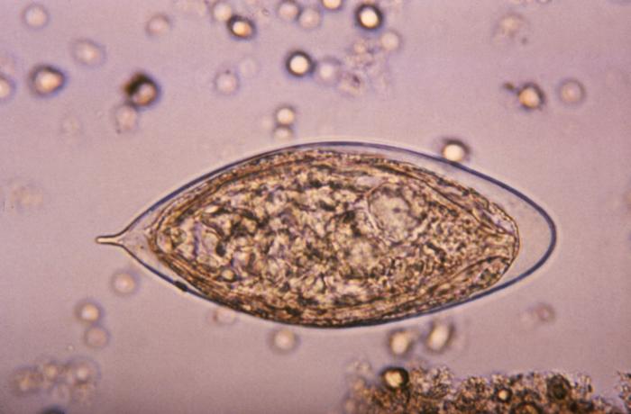
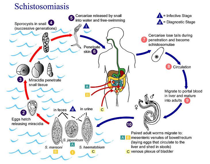
På Wikipedia kan man læse følgende om sygdommen bilharziose (tilgået d. 20. maj 2010):
Se mere udførlige beskrivelser af sygdommen på den engelske udgave af Wikipedia og på CDC / Division of Parasitic Diseases' side om Schistosomiasis. Marc Armand RufferNote on the presence of "Bilharzia haematobia" in Egyptian mummies of the Twentieth Dynasty (1250-1000 B.C.)(British Medical Journal, January 1, 1910) In a pirvious note published in this Jounual, I decscribed a process by which mummified tissues could be prepared for histological examination. I ventured to predict that it was highly probable that, by this method, one would be able to recognize pathological changes, such as cirrhosis, cancer, etc. Thanks to the kindness of Professor Elliot Smith, Professor Flinders Petrie, and Professor Keatinge, I have obtained several organs from mummies of the eighteenth to the twentieth dynasty, and I may state at once that such diseases as atheroma, pneumonia, renal abscesses, and cirrhosis of the liver are plainly recognizable. In the renal abscesses and in other lesions I have stained microorganisms with methylene blue, fuchsin, haematoxylin, and even by Gram's method. At the present time there is perhaps no disease more important to Egypt than that caused by the Bilharzia haematobia. So far no evidence has been produced to show how long it has existed in this country, although medical papyri contain prescriptions against one of its most prominent symptoms namely, haematuria. The lesions of this disease are best seen in the bladder and rectum, but unfortunately these are just the two mummified organs which I have not been able to obtain so far. Nevertheless, in the kidneys of two mummies of the twentieth dynasty, I have demonstrated in microscopic sections a large number of calcified eggs of Bilharzia haematobia, situated, for the most part, among the straight tubules. Although calcified, these eggs are easily recognizable and cannot be mistaken for anything else. I may add that I showed some of my sections to Professors Looss and Ferguson, whose paramount authority on such a subject cannot be disputed, and both confirmed my diagnosis. I have examined microscopically the kidneys of six mummies. The kidneys of two were apparently healthy; the left kidney of another was congenitally atrophied; those of the fourth contained multiple abscesses with well-staining bacteria and other lesions, which so far I have not diagnosed; those of the fifth and sixth showed Bilharzia eggs, and the latter had other lesions as well, which, owing to the shrunken state of the organ, I am unable to define accurately as yet.
|
|
© Copyright mag.art. Jan Helldén Præstegårdsvænget 30B 5210 Odense NV |
Hjem | Kommunikation og kognition | Medicinens historie | Biologisk antropologi | Blog |